

In a Nutshell
- The brain can reorganize and adapt at any age, not just in childhood, a fact scientists only fully accepted within roughly the past 50 years, after decades of assuming the adult brain was fixed.
- Children who have an entire half of their brain surgically removed to stop severe seizures can still recognize words and faces correctly more than 80% of the time using only the half that’s left, according to a 2022 study of 40 patients.
- Brain-training apps and games do produce real gains, but a review pooling 16 studies of over 1,500 older adults found the improvement is modest and mostly limited to the exact skill practiced, not everyday thinking or memory in general.
- A stroke rehabilitation technique that restrains a patient’s stronger arm to force use of the weaker one produces a real, moderate improvement in arm movement, but it helps walking and long-term recovery far less reliably.
- Whether adult human brains grow entirely new brain cells is a genuine, unresolved scientific debate: two major studies published in the same year, using similar tissue samples, reached opposite conclusions.
Neuroplasticity is the capacity of the brain to change its structure and activity in response to experience, learning, injury, or sensory input, at any point across the lifespan. The idea replaced an older assumption, held by most neuroscientists until the 1970s, that the adult brain’s wiring was essentially fixed after childhood. That assumption is now known to be wrong. The brain remains capable of forming new synaptic connections, strengthening or weakening existing ones, and in some regions and species generating new cells, well into old age.
What has changed since the 1970s is not whether the brain can adapt, but how much, where, and under what conditions, questions that remain active areas of research rather than settled fact.
What actually changes when a brain becomes more “plastic”?
Researchers generally split neuroplasticity into two broad categories. Structural plasticity refers to physical changes in brain anatomy: new dendritic spines (the small protrusions on neurons that receive signals from other cells), the sprouting of new axon branches, changes in grey matter volume, and, in some brain regions, the birth of entirely new neurons, a process called neurogenesis. Functional plasticity refers to the brain reassigning a cognitive job to a different network, either because a region was damaged or because repeated practice expanded the territory devoted to a skill.

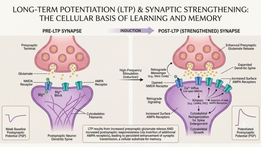
At the level of individual connections, the core mechanism is synaptic plasticity: synapses, the junctions between neurons, strengthen or weaken based on how they’re used. When repeated activity strengthens a connection, the effect is called long-term potentiation (LTP); when it weakens a connection, the effect is long-term depression (LTD). Both are well established in animal models and are the leading candidate mechanism for how memories are stored. A related process, homeostatic plasticity, keeps overall neural activity within a stable range so that learning-related changes don’t spiral out of control. None of this requires new neurons. Most plasticity, at any age, involves existing neurons adjusting the strength and pattern of their connections rather than the brain growing new cells.
Neurons are not the only cells involved. A 2024 study in Cell Reports, from researchers at Harvard Medical School, the University of Trento, and the German Center for Neurodegenerative Diseases, found that clusters of extracellular matrix molecules called chondroitin sulfates, structures more commonly associated with cartilage, coat groups of synapses in the mouse hippocampus and are necessary for synaptic plasticity and spatial memory. It’s one line of evidence, in mice, for a broader shift in the field toward treating non-neuronal cells as active participants in plasticity rather than passive support structures.
How did scientists conclude the brain could rewire itself?
The word “plasticity” was first applied to behavior by the psychologist William James in 1890, and the neuroanatomist Santiago Ramón y Cajal used a similar term around the same period to describe non-pathological change in the adult brain. Neither claim gained much traction. For most of the 20th century, the dominant view held that the brain’s structure was fixed once development ended.
That view was overturned by a series of experiments rather than a single discovery. In the 1960s, David Hubel and Torsten Wiesel found that sewing one eye of a kitten shut during early life caused the brain region tied to that eye to be taken over by the open eye, work that later earned them a Nobel Prize and established the concept of a critical period, a developmental window during which the brain is unusually receptive to being shaped by experience. Around the same time, the neuroscientist Michael Merzenich showed that adult monkeys’ brain maps of the hand could reorganize after a peripheral nerve was cut and allowed to regenerate, evidence that plasticity was not confined to childhood.
In the 1990s, neuroscientist Eleanor Maguire documented that licensed London taxi drivers, who memorize the city’s street layout for a rigorous qualifying exam, had measurably larger posterior hippocampi than non-drivers, one of the most widely cited pieces of evidence for structural plasticity from real-world experience in adult humans. Merzenich shared the 2016 Kavli Prize in Neuroscience with Eve Marder and Carla Shatz for their combined contributions to understanding how experience remodels the brain, from individual synapses up to whole cortical regions, as the three laureates discussed in a joint interview around the prize.
Why is a child’s brain more plastic than an adult’s?
Plasticity is not switched off in adulthood, but it is dialed down, and understanding why has become one of the more active corners of the field. A December 2025 study from the Salk Institute, published in Nature, identified a protein called CCN1, secreted by star-shaped support cells called astrocytes, as a molecular brake that stabilizes neural circuits in the adult mouse visual cortex. Removing CCN1 in adult mice destabilized circuits that are normally locked in place, while boosting it during the developmentally plastic critical period made circuits mature and stabilize early. The finding is preclinical, in mice, and has not yet been tested in humans, but it adds mechanistic detail to a broader pattern: the adult brain appears to actively suppress plasticity in already-tuned circuits rather than simply losing the capacity for it, which raises the possibility that pharmacologically loosening that brake could, in principle, reopen windows of plasticity later in life.
This matters clinically because certain functions, such as binocular depth perception or first-language phonology, were long thought to require exposure during a narrow childhood window or be lost permanently. That absolute view has softened. Adults with amblyopia (a vision disorder sometimes called lazy eye) and other stereo-vision deficits have shown genuine, if partial, improvement with targeted training well outside the classic critical period, an active area of ophthalmological and neuroscience research rather than settled treatment.
Can the brain really compensate for major injury or missing tissue?
The clearest human evidence for the brain’s capacity to reroute function comes from children who undergo hemispherectomy, the surgical removal or disconnection of an entire brain hemisphere, most often to control severe, drug-resistant epilepsy. A 2022 study from the University of Pittsburgh and Carnegie Mellon University, published in the Proceedings of the National Academy of Sciences, tested 40 hemispherectomy patients on word and face recognition, tasks that in an intact brain are typically split between the left and right hemispheres. The single remaining hemisphere supported both functions, with accuracy exceeding 80% and a gap of less than 10 percentage points compared with matched controls. The result does not mean the brain fully compensates: the study’s own authors describe the remaining hemisphere’s performance as adequate but suboptimal, and the degree of recovery in any individual case cannot be predicted in advance.
A related line of evidence comes from cross-modal plasticity, the reorganization of a sensory brain region deprived of its usual input to process a different sense. A 2026 study in Human Brain Mapping, led by researchers affiliated with the universities of Coimbra, Glasgow, Padua, and Peking, used functional MRI in congenitally deaf and hearing individuals viewing simple visual patterns. In deaf participants, the auditory cortex, which normally processes sound, showed an organized, location-specific pattern of deactivation in response to visual stimuli, a mechanism the authors interpret as the brain filtering or encoding visual information through a region deprived of its original input, distinct from the more commonly described pattern of increased cross-modal activation. It is a single imaging study and a novel finding within the field, so it should be read as adding nuance to, not overturning, the existing evidence for cross-modal reorganization in deafness and blindness.
For adults recovering from stroke, the best-tested rehabilitation approach grounded in plasticity is constraint-induced movement therapy, which restrains the unaffected limb to force intensive use of the affected one. Cochrane-reviewed trials and subsequent meta-analyses find a moderate, statistically significant improvement in arm motor activity (standardized mean difference around 0.51) and a smaller effect on arm motor function (around 0.28) in the months after treatment. Effects on lower-limb outcomes such as gait and balance are considerably less consistent across trials, and one trial sequential analysis has cautioned that even the positive upper-limb findings may not yet have accumulated enough data to rule out a false positive. Constraint-induced therapy is one of the more rigorously tested plasticity-based interventions in rehabilitation medicine, not proof that plasticity-based approaches uniformly work. Cognitive, rather than physical, rehabilitation shows a similar pattern of modest but real change: a small randomized study of 17 adults with chronic traumatic brain injury, published in the Journal of Neurotrauma, found that 40 sessions of computerized cognitive training over 14 weeks measurably altered white matter microstructure, changes that tracked with improvements in processing speed, attention, and working memory. The sample is small enough that the finding should be treated as promising rather than conclusive.
Does commercial “brain training” actually work?
This is where the evidence and the marketing claims diverge most sharply. In 2014, two groups of scientists, one with more than 70 signatories and one with 133, published dueling public statements reaching opposite conclusions about whether commercial brain-training software meaningfully improves cognition or delays decline. In 2016, the U.S. Federal Trade Commission required Lumos Labs, maker of the Lumosity app, to pay $2 million to settle charges that it made unsupported claims linking its games to reduced risk of dementia and Alzheimer’s disease, and ordered the company to hold competent clinical evidence before making similar claims again.
The evidence since then supports a narrower, more modest picture. A meta-analysis of 16 randomized controlled trials covering 1,543 older adults without cognitive impairment found statistically significant but modest improvements from commercial computerized cognitive games: a standardized mean difference of 0.40 for processing speed, 0.21 for working memory, 0.21 for executive function, and 0.12 for verbal memory, with no significant effect on attention or visuospatial ability.
The consistent caveat across this literature is transfer: gains are strongest on tasks that resemble the training itself (near transfer) and weaken substantially for broader, real-world cognitive benefits (far transfer). A single randomized trial from McGill University, published in 2025 in JMIR Serious Games, reported that 10 weeks of the commercial program BrainHQ increased a marker of cholinergic brain health, measured with PET imaging, in 92 healthy older adults to a level the researchers likened to that typical of someone roughly a decade younger. It’s a striking single-study finding, conducted with the training software’s maker providing product access while the university team ran the trial independently, and it awaits replication before it can be treated as established.
Do drugs, exercise, or other interventions promote plasticity?
Several categories of intervention have credible, if uneven, evidence behind them. Aerobic exercise is among the best supported: consistent evidence links it to increased volume in the hippocampus and improvements in memory-related tasks in both animal models and human trials. Meditation practice has been associated with measurable differences in cortical thickness in attention-related regions, most notably in studies led by Sara Lazar at Harvard, though sample sizes in this literature tend to be small.
Pharmacologically, the most consequential development of the past decade has been the recognition that fast-acting antidepressants such as ketamine work partly by rapidly increasing dendritic spine density and restoring synaptic connectivity, in contrast to traditional antidepressants, which also promote plasticity but over a much slower timescale of weeks to months. That mechanistic insight has driven a wave of research into other compounds that promote plasticity through similar pathways, now sometimes called psychoplastogens, including classic and novel psychedelics.
Most of this evidence remains preclinical. A 2025 mouse study published in the journal Psychedelics found that a single dose of a psychedelic compound improved cognitive flexibility in a reversal-learning task for two to three weeks afterward; it is an animal study, and its authors themselves note that basic questions, such as what happens with repeated dosing, remain unanswered. A non-hallucinogenic compound called zalsupindole, developed by a venture-backed biotechnology company and profiled in a 2026 review in ACS Chemical Neuroscience, has shown plasticity-promoting effects comparable to ketamine and psilocybin in animal studies and has completed an early Phase 1 safety trial in humans; it is not an approved treatment and remains in early clinical development for major depressive disorder.
Other approaches target the timing rather than the chemistry of learning. A 2022 study from the University of Cambridge, published in Cerebral Cortex, found that briefly matching a visual learning task to each of 80 participants’ individual brainwave rhythm, measured by EEG, tripled their rate of improvement on a subsequent cognitive task compared with mismatched or random timing, an effect that persisted when participants were retested the next day. It is one study, but it points to a broader and still-developing idea that the brain’s receptivity to new information fluctuates on a rhythmic, and possibly circadian, basis, a pattern also observed in a 2025 rat study from Tohoku Universityshowing that the brain’s capacity for long-term potentiation shifted across the day in step with the animals’ sleep-wake cycle.
Does plasticity decline with age, and is that entirely bad?
Yes, on average, but unevenly and not as steeply as once assumed. Gene-expression studies of human frontal cortex tissue show reduced expression, after around age 40 and more sharply after 70, of genes central to synaptic plasticity, alongside a rise in oxidative DNA damage. A 2025 study from DZNE, the University of Magdeburg, and the University of Tübingen, published in Nature Neuroscience, used ultra-high-resolution 7 Tesla MRI to scan the primary somatosensory cortex, the brain region that processes touch, in roughly 60 adults aged 21 to 80. It found that cortical thinning with age was not uniform across the tissue’s layers: the layers that receive the most continuous sensory stimulation stayed stable or even thickened, while less-used deeper layers thinned, though those deeper layers showed a partial compensatory rise in myelin content, a substance that speeds nerve signal transmission. The finding is observational, drawn from cross-sectional brain scans rather than a study that followed the same people over time, so it shows an association between activity and preserved cortical structure rather than proof that stimulation itself caused the preservation.
What don’t we know yet?
Several open questions sit underneath the plasticity research described above. The most consequential is whether the adult human hippocampus produces new neurons at all. Two studies published in 2018, using similar postmortem tissue and staining techniques, reached opposite conclusions: one reported that neurogenesis becomes undetectable after adolescence, the other reported that it persists throughout life at a declining rate. A widely cited 2018 review in Cell Stem Cell argued there was no reason to abandon the idea that adult-generated neurons matter functionally, but the field has not converged, and more recent transcriptomic work has continued to find the signature of neurogenesis in adult mice, macaques, and pigs but not consistently in adult humans.
Beyond that central debate, more than 130 clinical trials registered on ClinicalTrials.gov currently reference neuroplasticity as a study condition or mechanism, spanning spinal cord injury rehabilitation, post-concussive cognitive symptoms, schizophrenia-related brain stimulation, and cognitive training in mild cognitive impairment, an indication of how much of this field is still being tested rather than applied. Whether astrocyte-secreted molecules such as CCN1 behave the same way in the human brain as in mice is untested. Whether non-hallucinogenic psychoplastogens will match ketamine’s clinical effect in controlled human trials, rather than animal models, is unknown. And whether brainwave-timed learning techniques generalize beyond simple visual tasks to real classrooms and training environments has not yet been established.
Frequently Asked Questions
Is neuroplasticity the same thing as “rewiring your brain”?
Loosely, yes, but the popular phrase overstates how dramatic or intentional most plasticity is. Most changes involve existing neurons strengthening, weakening, or slightly restructuring their connections in response to repeated experience, not large-scale rerouting. Dramatic reorganization, such as one brain hemisphere taking over functions normally split between two, occurs mainly after significant injury, most reliably in children.
Can adults still learn a new language as easily as children?
Adults generally learn additional languages more slowly and with a stronger accent than children who acquire them early, particularly for pronunciation. But adult language learning still produces measurable structural brain changes, and functional fluency remains achievable at any age; it typically requires more deliberate, sustained practice than it does in early childhood.
Do brain-training apps prevent dementia?
There is no reliable evidence that commercial brain-training apps prevent or delay dementia or Alzheimer’s disease. Regulators penalized at least one major brain-training company for making such claims without adequate evidence. Modest, task-specific cognitive gains from computerized training are supported by meta-analysis, but broad protective claims are not.
Does exercise really change the brain, or is that oversold?
Aerobic exercise has some of the more consistent evidence behind it among plasticity-promoting interventions, with links to hippocampal volume and memory performance across animal and human studies. It is not a cure for cognitive decline, but among lifestyle interventions it has one of the stronger evidence bases.
What is a “critical period” and does missing one mean permanent loss?
A critical period is a developmental window during which a brain circuit is unusually sensitive to being shaped by experience, the classic example being early visual development. Missing a critical period can produce lasting deficits, such as amblyopia if not treated in early childhood, but “permanent” has proven less absolute than once believed: some adults with long-standing visual or sensory deficits show partial improvement with targeted, intensive training later in life.
Does the brain create new neurons in adulthood?
This remains scientifically unresolved for the human hippocampus specifically. Adult neurogenesis is well established in several other mammal species. In humans, two major 2018 studies using similar methods reached opposite conclusions, and the question has not been definitively settled since.
- Chelini G, Mirzapourdelavar H, Durning P, et al. Focal clusters of peri-synaptic matrix contribute to activity-dependent plasticity and memory in mice. Cell Reports. 2024. DOI: 10.1016/j.celrep.2024.114112. Animal (mouse) study.
- Fracasso A, et al. The Neural Organization of Visual Information in the Auditory Cortex of the Congenitally Deaf. Human Brain Mapping. 2026. DOI: 10.1002/hbm.70444. Human imaging study.
- Voelbel G, et al. Computerized Cognitive Remediation Affects White Matter Microstructure in Relation to Improved Cognitive Function in Adults with Chronic Traumatic Brain Injury. Journal of Neurotrauma. 2026. DOI: 10.1177/089771512514140. Randomized study, n=17.
- Sancho L, Allen N, et al. Astrocyte CCN1 stabilizes neural circuits in the adult brain. Nature. 2025. DOI: 10.1038/s41586-025-09770-w. Animal (mouse) study.
- Attarha M, de Villers-Sidani E, et al. Effects of Computerized Cognitive Training on Vesicular Acetylcholine Transporter Levels… Results from the INHANCE Randomized Clinical Trial. JMIR Serious Games. 2025. DOI: 10.2196/75161. RCT, n=92.
- The Kavli Foundation. Interview with 2016 Kavli Prize in Neuroscience laureates Eve Marder, Michael Merzenich, and Carla Shatz. 2016. kavliprize.org.
- Brouns EJ, Ekins T, Ahmed OJ. Single-dose psychedelic enhances cognitive flexibility and reversal learning in mice weeks after administration. Psychedelics. 2025. DOI: 10.61373/pp025r.0002. Animal (mouse) study.
- Kühn E, et al. Layer-specific changes in sensory cortex across the lifespan in mice and humans. Nature Neuroscience. 2025. DOI: 10.1038/s41593-025-02013-1. Observational, human n≈60 plus mouse data.
- Donen Y, Ikoma Y, Matsui K. Diurnal modulation of optogenetically evoked neural signals. Neuroscience Research. 2025. DOI: 10.1016/j.neures.2025.104981. Animal (rat) study.
- Michael E, Kourtzi Z, Leong V, et al. Learning at your brain’s rhythm: individualized entrainment boosts learning for perceptual decisions. Cerebral Cortex. 2022. DOI: 10.1093/cercor/bhac426. RCT-style study, n=80.
- Granovetter M, Robert S, Ettensohn L, Behrmann M. With childhood hemispherectomy, one hemisphere can support, but is suboptimal for, word and face recognition. PNAS. 2022. DOI: 10.1073/pnas.2212936119. Human cohort, n=40.
- Sorrells SF, Paredes MF, Cebrian-Silla A, et al. Human hippocampal neurogenesis drops sharply in children to undetectable levels in adults. Nature. 2018. DOI: 10.1038/nature25975.
- Boldrini M, Fulmore CA, Tartt AN, et al. Human Hippocampal Neurogenesis Persists throughout Aging. Cell Stem Cell. 2018. DOI: 10.1016/j.stem.2018.03.015. These two studies used similar postmortem methods and reached opposite conclusions.
- Kempermann G, et al. Human Adult Neurogenesis: Evidence and Remaining Questions. Cell Stem Cell. 2018. DOI: 10.1016/j.stem.2018.04.004. Review.
- Simons DJ, et al. Do “Brain-Training” Programs Work? Psychological Science in the Public Interest. 2016. PMID: 27697851. Systematic review.
- Meta-analysis: The use of commercial computerised cognitive games in older adults. Scientific Reports. 2020. DOI: 10.1038/s41598-020-72281-3. Meta-analysis of 16 RCTs, n=1,543.
- U.S. Federal Trade Commission. Lumosity to Pay $2 Million to Settle FTC Deceptive Advertising Charges for Its “Brain Training” Program. 2016. ftc.gov.
- A meta-analysis of constraint-induced movement therapy after stroke. Journal of Rehabilitation Medicine. 2014. medicaljournalssweden.se. Meta-analysis.
- Constraint-induced movement therapy: trial sequential analysis applied to Cochrane collaboration systematic review results. 2015. PMC4307139.
- Salfiti M, Kyriazis M, Mikellides G. Zalsupindole: A Non-Hallucinogenic Psychoplastogen Advancing Psychedelic-Inspired Therapeutics. ACS Chemical Neuroscience. 2026. PMID: 41493772. Review; sponsor-affiliated development program, Phase 1 data in humans.
- ClinicalTrials.gov. Search results for studies referencing “neuroplasticity,” accessed August 2026. clinicaltrials.gov.


Cite This Page